合成ヒト胚モデルの利用法

2023年9月8日
全体に公開

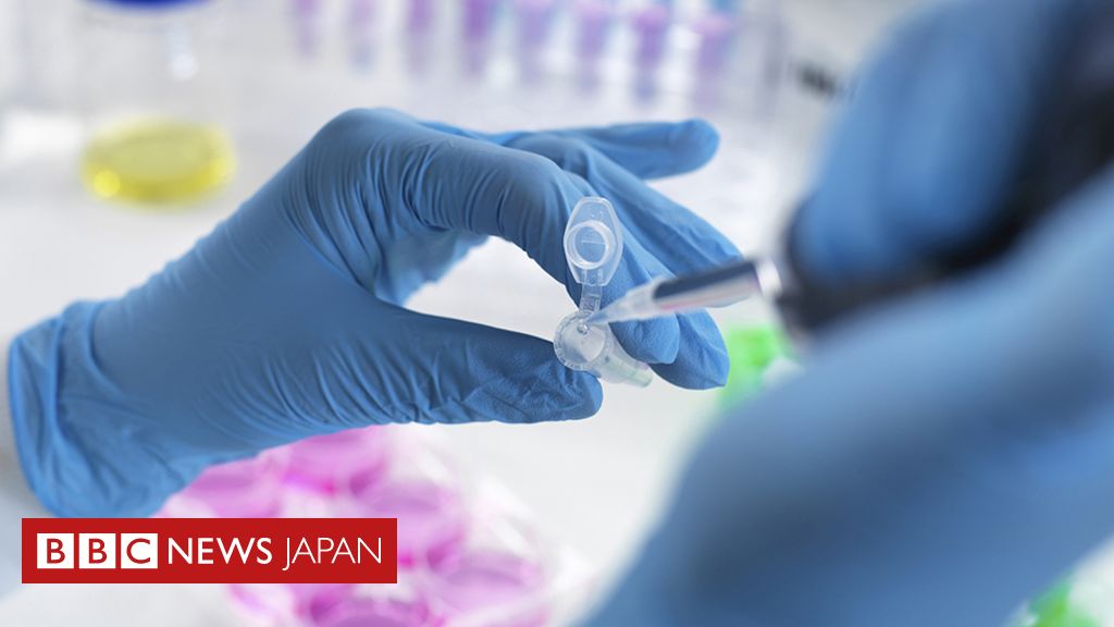
イスラエル・ワイツマン科学研究所のJacob Hanna博士の研究チームが、実験室で培養した幹細胞からヒト胚のモデルを作成し、子宮の外で14日目まで成長させることに成功しました。この合成胚モデルは、胎盤、卵黄嚢、絨毛膜嚢、さらに他の外部組織を含む、この段階に特徴的なすべての構造を持っているということです。

9月7日、BBCの英語ニュースを聴いていたら、トップで報道していたニュースですが、日本語ニュースにもなっています。

Oldak, B. et al. (2023) Complete human day 14 post-implantation embryo models from naïve ES cells. Nature https://doi.org/10.1038/s41586-023-06604-5

Complete human day 14 post-implantation embryo models from naïve ES cells - Nature
The ability to study human post-implantation development remains limited due to ethical and technical challenges associated with intrauterine development after implantation1. Embryo-like models with spatially organized morphogenesis of all defining embryonic and extra-embryonic tissues of the post-implantation human conceptus (i.e., embryonic disk, bilaminar disk, yolk- and chorionic sacs, surrounding trophoblasts) remain lacking2. Mouse naïve embryonic stem cells (ESCs) have recently been shown to give rise to embryonic and extra-embryonic stem cells capable of self-assembling into post-gastrulation mouse Structured Stem cell-based Embryo Models with spatially organized morphogenesis (SEMs)3. Here, we extend these findings to humans, while using only genetically unmodified human naïve ESCs (in HENSM conditions)4. Such human fully integrated SEMs recapitulate the organization of nearly all known lineages and compartments of post-implantation human embryos including epiblast, hypoblast, extra-embryonic mesoderm, and trophoblast surrounding the latter layers. These human complete SEMs demonstrated developmental growth dynamics that resemble key hallmarks of post-implantation stage embryogenesis up to 13-14 days post-fertilization (dpf) (Carnegie stage 6a). This includes embryonic disk and bilaminar disk formation, epiblast lumenogenesis, polarized amniogenesis, anterior-posterior symmetry breaking, PGC specification, polarized yolk sac with visceral and parietal endoderm, extra-embryonic mesoderm expansion that defines a chorionic cavity and a connecting stalk, a trophoblast surrounding compartment demonstrating syncytium and lacunae formation. This SEM platform may enable the experimental interrogation of previously inaccessible windows of human early post-implantation up to peri-gastrulation development.
www.nature.com

実は、この論文、去年の秋には、bioRxivにプレプリントとして掲示されていましたので、査読後、雑誌掲載で改めてニュースになったという感じです。タイトルや文章が少し変わっていますが、Nature誌の場合、編集過程で変更されることがしばしばあります。

Transgene-Free Ex Utero Derivation of A Human Post-Implantation Embryo Model Solely from Genetically Unmodified Naïve PSCs
Our ability to study early human post-implantation development remains highly limited due to the ethical and technical challenges associated with intrauterine development of the human embryo after implantation. Despite the great progress made on human gastruloids, axioloids and in vitro cultured blastoids, such elegant models do not constitute an integrated Stem cell-derived Embryo Models (SEMs) that includes all the key extra-embryonic tissues of the early post-implantation human conceptus (e.g., hypoblast, yolk-sac, trophoblasts, amnion, and extraembryonic mesoderm), and thus, do not recapitulate post-implantation epiblast development within the context of these extra-embryonic compartments. Mouse naïve pluripotent stem cells (PSCs) have recently been shown to give rise to embryonic and extra-embryonic stem cells capable of self-assembling into post-gastrulation mouse SEMs, while bypassing the blastocyst-like stage, and eventually initiating organogenesis ex utero . Here, we implement critical adaptations to extend these finding to humans, while using only genetically unmodified human naïve PSCs, thus circumventing the need for ectopic expression of lineage promoting transgenes. Such integrated human SEMs recapitulate the organization of all known compartments of early post-implantation stage human embryos, including epiblast, hypoblast, extra-embryonic mesoderm, and trophoblast surrounding the latter layers. The organized human SEMs recapitulate key hallmarks of post-implantation stage embryogenesis up to 13-14 days post-fertilization (dpf, Carnegie stage 6a), such as bilaminar disk formation, epiblast lumenogenesis, amniogenesis, anterior-posterior symmetry breaking, PGC specification, primary and secondary yolk sac formation, and extra-embryonic mesoderm expansion that defines a chorionic cavity and a connective stalk. This new platform constitutes a tractable stem cell-based model for experimentally interrogating previously inaccessible windows of human peri- and early post-implantation development. ### Competing Interest Statement Declaration of interests: J.H.H has submitted patent applications relevant to the findings reported herein and is a chief scientific adviser of Renewal Bio Ltd. that has licensed technologies described herein and sponsored some parts of this project.
www.biorxiv.org

既にマウスを使っての応用研究は実施されており、こういう記事のソースにもなっています。

幹細胞から人工胚、「最高の臓器プリンター」目指すイスラエル企業

RenewalBioというのが、このイスラエル企業です。

また、別のグループも昨年、同様な学会発表を行っており、その報道がありました。

BBCの報道では、このような合成胚モデルの利用法について、以下のように説明しています。

胚モデルについては、科学者たちがさまざまな種類の細胞がどう出現するのかを説明したり、体の器官を作る最も初期の段階を観察したり、遺伝性の疾患について理解を深めたりすることに役立つことが期待されている。

また、このような合成胚モデルは、移植組織や臓器を作るための新しい方法に利用できる可能性があります。さらに、本物の胚では実行できない実験、たとえば薬物やその他の物質への曝露が胎児の発育に与える影響を調べることができるようになるかもしれません。

幹細胞から作った分化細胞を用いる様々な研究や治療法の開発は既にいろいろな形で行われてきていますが、これまでとは違った種類の細胞や組織を作ったりすることができるようになるということです。

この研究を行っているJacob Hanna博士は、上のTechnology Reviewの記事では、「妊娠40日から50日に相当するヒト胚の人工モデル」が目標とも語っています。本物のヒト胚の場合、14日以上のヒト胚をどう扱うのかという倫理的な問題が関わってきます。

しかし、今回の胚モデルは、幹細胞から作ったモデルであって、本物のヒト胚ではありません。

一方、幹細胞から脳のような組織を作る「ヒト脳オルガノイド」、ヒト細胞を実験動物に移植して脳組織を作らせた場合、「意識」を持つ可能性なども議論されています。

「妊娠40日から50日に相当するヒト胚の人工モデル」から、移植に使える細胞を採取して治療に利用するということをどう考えたらよいのでしょうか。カズオ・イシグロの「Never Let Me Go」みたいな世界はあまり想像したくないです。

合成生物学は新たな産業革命の鍵となるか?」担当:山形方人

【Twitter】 https://twitter.com/yamagatm3

応援ありがとうございます!
いいねして著者を応援してみませんか


このトピックスについて
津覇 ゆういさん、他1329人がフォローしています